Academic Centre
The Human Pathology Collection of the UL Museum
After the founding of the University of Latvia in 1919, an experienced pathologist, Professor Roman Adelheim (1881-1938), was entrusted with organizing the training of students in Pathology. He established the Department of Pathological Anatomy, set up student auditoriums, a pathohistological laboratory, and a library in 1921, and laid the foundation for the Museum of Pathological Anatomy.
When the Faculty of Medicine was reopened at the University of Latvia in 1997 after a break of 47 years, the Museum of Human Pathology also resumed its work.
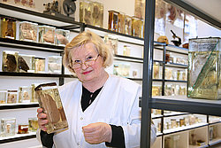
The Human Pathology Collection of the Museum of the University of Latvia provides UL students and resident doctors of various specialties with material that allows them to visualize the changes in the body caused by diseases as well as to learn the pathomorphology and pathogenesis of diseases in depth.
The collection includes a large number of macropreparations of pathologically altered human organs, micropreparations, moulages of pathologically altered organs and posters that show a variety of pathological changes in the human body at the macroscopic and microscopiclevel.
The 2nd, 3rd and 4th year students of the Faculty of Medicine, as well as resident doctors are provided with the Museum's visual material for studyinh Pathology and other study coursespic to be studied every week.
The museum offers everyone to get acquainted with various pathologies of the human heart and blood vessels, liver, kidney, brain, skin, intestinal and other localization pathologies, macro-preparations of human embryos and fetuses at different stages of development, as well as a rich collection of stones found in several organs and a well preserved collection of micropreparations and other interesting objects from 1897.
The museum offers:
- to get acquainted with the historical overview of the development of Pathology at the University of Latvia;
- a tour through the collection on the most common pathologies in Latvia and in the world;
- interactive lessons in the microscpy laboratory on the differences in the structure of pathologically altered and normal cells.
Expert